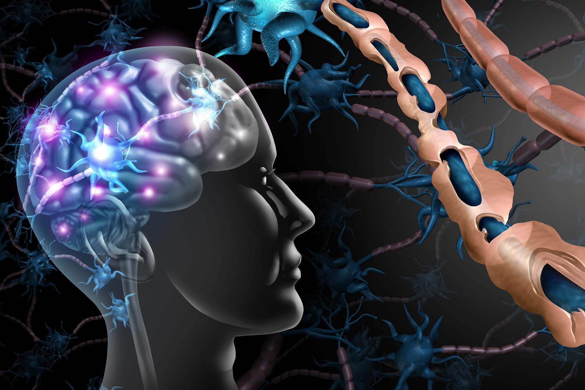
It’s there.
It has always been there.
Standard MRI scans missed it.
Clinicians looked right past it.
For decades, multiple sclerosis research operated with half the picture. White matter damage shows up clearly. You can see it. You can count it. But the real troublemaker—the kind of lesion tied directly to disability and the slow creep of cognitive decline—lurked in the gray matter. Specifically, the cortex.
Invisible to the naked eye on routine scans.
This left doctors blind. Not literally, obviously. But clinically? Yeah, blind.
Current MS drugs mostly target white matter. That leaves the cortical lesions unchecked. And that matters because those hidden injuries drive the disease forward. We knew they existed, thanks to post-mortem tissue studies, but we couldn’t see them in living patients.
Now, artificial intelligence fixes that gap.
The AI Difference
A team led by the University at Buffalo trained an algorithm to look deeper. Much deeper.
Published in Communications Medicine, the study details how computational methods compare data across multiple images. One image doesn’t show much. Put three or four together? The AI finds the pattern. It pulls the disease signals out of the noise that conventional viewing misses entirely.
“Detecting previously invisible cortical lesions… has major implications,” says senior author Robert Zivadinov.
He isn’t just talking about cool tech. He’s talking about seeing, for the first time, the actual drivers of MS progression in standard legacy scans.
Michael G. Dwyer knows the frustration. First author of the paper and a neurologist himself.
“We have all been very frustrated,” Dwyer says.
Frustrated because histopathologists had proven for decades that these lesions were wrecking brain tissue. We just couldn’t prove it on a living patient’s MRI. Until now.
“There’s a lot of ongoing that continues to happen… you won’t see with conventionalMRI, but that histopathologists have clearly demonstrated… for decades.”
The AI doesn’t invent data. It synthesizes what is missing. It looks at minor discrepancies between contrast images that a human eye—or even a standard software filter—would ignore.
11,000 Missed Lesions
The test case was robust.
ORATORIO.
A massive Phase III clinical trial for the MS drug Ocrevizumab.
700+ participants.
Standard MRIs.
The researchers applied their new multimodal cortical lesion enhancement method, dubbed MMCLE.
The result?
Standard scans showed white matter issues.
AI-guided processing revealed a hidden layer of devastation.
About 15 to 20 new lesions per patient.
Across the entire dataset? More than 11,000 previously hidden lesions detected.
Did the radiologists miss them on purpose?
No.
They were literally invisible without the computational assist.
Dwyer points out the power of generative AI here. It spots tissue behaving “wrong” by comparing it across different contrast layers. Healthy tissue acts one way. Damaged cortex acts another. The AI spots the mismatch.
This matters because the ORATORIO data is historical. It was already collected. It was “finished.”
But Zivadinov suggests this work changes how we review all that data.
It also changes how we design future trials.
Genentech helped support the work. No surprise. They make the drug being tested.
The collaboration combined academic rigor with industrial muscle.
So we finally have the full map.
Or do we?
The blind spot is gone.
But what will doctors do with all this new damage they can suddenly see?
Will treatments shift to target the cortex?
Will the definition of “stable disease” change?
The scans are sitting right there.
We just finally learned how to look.